Proliferative enteropathy (proliferative enteritis, proliferative ileitis, intestinal adenomatosis) has been reported in several animal species including the pig,1 2 dog, 2 foal, 3 blue fox, 5 guinea pig, 4 ferret, 6 hamster, 7 and...
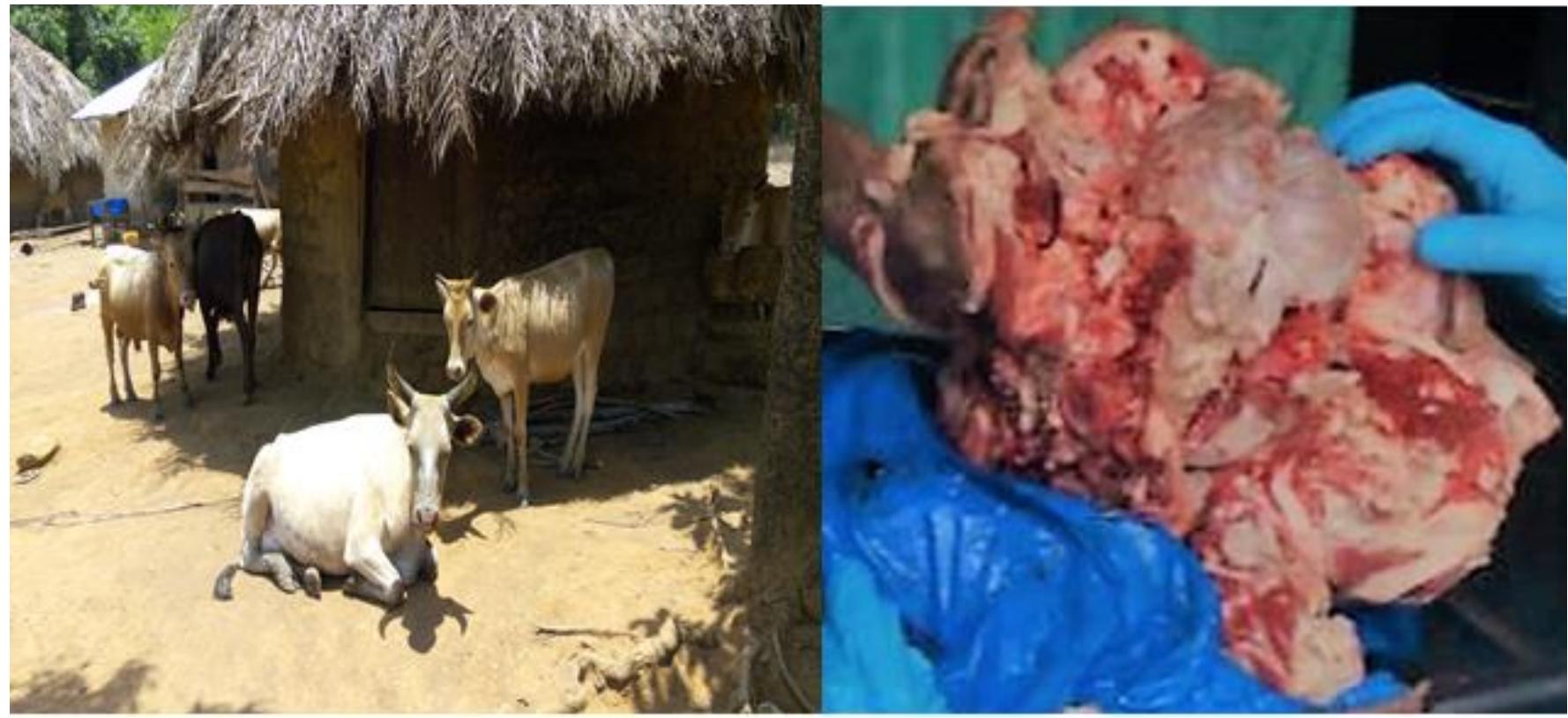
moreProliferative enteropathy (proliferative enteritis, proliferative ileitis, intestinal adenomatosis) has been reported in several animal species including the pig,1 2 dog, 2 foal, 3 blue fox, 5 guinea pig, 4 ferret, 6 hamster, 7 and rabbit. 13 The disease is characterized by adenomatous hyperplasia of crypt epithelial cells in the ileum and colon with intracytoplasmic curved bacteria resembling Campylobacter species. In the single case previously reported in a foal, Campylobacter-like organisms were demonstrated within the cytoplasm of enterocytes by spirochete stains and electron microscopy. In a study using cloned DNA probes to isolated Campylobacter-like organisms, there was failure of the probes to hybridize with common porcine Campylobacter species, suggesting the causative agent to be an unidentified or uncultured species. 8 This finding, along with DNA sequencing and structural characteristics, resulted in this organism being named ileal symbiont intracellularis. 9 Subsequently, the organism was described and classified as a new genus and species, Lawsonia intracellularis. 1 1 An unweaned 5-month-old mixed-breed female foal with a history of anorexia, lethargy, and profuse watery diarrhea of greater than 1-week duration was presented to the University of Kentucky, Livestock Disease Diagnostic Center, for necropsy. Treatment had not been attempted. At necropsy, the foal was thin with readily apparent skeletal muscle atrophy. Gross lesions were confined to the small intestine. There was irregular thickening of the jejunum and ileum with the ileum being more severely affected. Lesions in the midjejunum were multifocal in nature and consisted of areas of discoid mucosal thickening, whereas the distal jejunum and ileum contained diffuse mucosal thickening resulting in a rugose pattern (Fig. ). Samples of the small intestine, stomach, cecum, colon, brain, heart, lung, kidney, and spleen were placed in 10% neutral buffered formalin and, following fixation, were embedded in paraffin, sectioned at 5 µm, and stained with hematoxalin and eosin. In addition, selected sections of small intestine were stained by the Warthin-Starry silver impregnation method for bacteria. Pieces of formalinfixed small intestine were postfixed in osmium tetroxide and embedded in epoxy resin. Thin sections were stained with uranyl acetate and lead citrate. Histopathologically, the affected mucosa was thickened and consisted of hyperplastic glandular structures lined by immature epithelial cells (Fig. ). The hyperplastic epithelium resulted in distortion of the normal villous structure. Some From the Livestock Disease Diagnostic Center, University of Kentucky, 1429 Newtown Pike, Lexington, KY 40511 (Williams, Harrison), and the