Papers by Pierre-Frédéric Villard
Studies in health technology and informatics, 2009
We present an integrated system for training ultrasound guided needle puncture. Our aim is to pro... more We present an integrated system for training ultrasound guided needle puncture. Our aim is to provide a cost effective and validated training tool that uses actual patient data to enable interventional radiology trainees to learn how to carry out image-guided needle puncture. The input data required is a computed tomography scan of the patient that is used to create the patient specific models. Force measurements have been made on real tissue and the resulting data is incorporated into the simulator. Respiration and soft tissue deformations are also carried out to further improve the fidelity of the simulator.
International Journal of Computer Assisted Radiology and Surgery, 2014
Purpose Training in Interventional Radiology currently uses the apprenticeship model, where clini... more Purpose Training in Interventional Radiology currently uses the apprenticeship model, where clinical and technical skills of invasive procedures are learnt during practice in patients. This apprenticeship training method is increasingly limited by regulatory restrictions on working hours, concerns over patient risk through trainees' inexperience, and the variable exposure to case mix and emergencies during training. To address this, we have developed a computer-based simulation of visceral needle puncture procedures.
Developing an immersive ultrasound guided needle puncture simulator
Studies in health technology and informatics, 2009
We present an integrated system for training ultrasound guided needle puncture. Our aim is to pro... more We present an integrated system for training ultrasound guided needle puncture. Our aim is to provide a cost effective and validated training tool that uses actual patient data to enable interventional radiology trainees to learn how to carry out image-guided needle puncture. The input data required is a computed tomography scan of the patient that is used to create the patient specific models. Force measurements have been made on real tissue and the resulting data is incorporated into the simulator. Respiration and soft tissue deformations are also carried out to further improve the fidelity of the simulator.
Proceedings on Seventh International Conference on Information Visualization, 2003. IV 2003., 2000

Studies in health technology and informatics
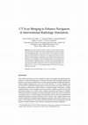
We present a method to merge two distinct CT scans acquired from different patients such that the... more We present a method to merge two distinct CT scans acquired from different patients such that the second scan can supplement the first when it is missing necessary supporting anatomy. The aim is to provide vascular intervention simulations with full body anatomy. Often, patient CT scans are confined to a localised region so that the patient is not exposed to more radiation than necessary and to increase scanner throughput. Unfortunately, this localised scanning region may be limiting for some applications where surrounding anatomy may be required and where approximate supporting anatomy is acceptable. The resulting merged scan can enhance body navigation simulations with X-ray rendering by providing a complete anatomical reference which may be useful in training and rehearsal. An example of the use of our CT scan merging technique in the field of interventional radiology is described.
International Conference on Medical Information Visualisation - BioMedical Visualisation (MediVis 2007), 2007
To monitor a lung mechanical model and then predict tumour motion we proposed a approach based on... more To monitor a lung mechanical model and then predict tumour motion we proposed a approach based on the pleura physiology. By comparing the predictions to landmarks set by medical experts, we observed better results with regards to the one obtained with approaches found in the literature. Beside, we focus on the rib cage kinematics, which play a significant role in the pleura outer-surface motion and therefore in the lung motion. We proposed a kinematic model of the rib cage based on the finite helical axis method and we show out interesting results.
Studies in health technology and informatics, 2011
Inguinal hernia repair procedures are often one of the first surgical procedures faced by junior ... more Inguinal hernia repair procedures are often one of the first surgical procedures faced by junior surgeons. The biggest challenge in this procedure for novice trainees is understanding the 3D spatial relations of the complex anatomy of the inguinal region, which is crucial for the effective and careful handling of the present anatomical structures in order to perform a successful and lasting repair. Such relationships are difficult to illustrate and comprehend through standard learning material. This paper presents our work in progress to develop a simulation-based teaching tool allowing junior surgeons to train the Lichtenstein tension-free open inguinal hernia repair technique for direct and indirect hernias, as well as to enforce their understanding of the spatial relations of the involved anatomy.
Studies in health technology and informatics, 2009
We present a method to merge two distinct CT scans acquired from different patients such that the... more We present a method to merge two distinct CT scans acquired from different patients such that the second scan can supplement the first when it is missing necessary supporting anatomy. The aim is to provide vascular intervention simulations with full body anatomy. Often, patient CT scans are confined to a localised region so that the patient is not exposed to more radiation than necessary and to increase scanner throughput. Unfortunately, this localised scanning region may be limiting for some applications where surrounding anatomy may be required and where approximate supporting anatomy is acceptable. The resulting merged scan can enhance body navigation simulations with X-ray rendering by providing a complete anatomical reference which may be useful in training and rehearsal. An example of the use of our CT scan merging technique in the field of interventional radiology is described.
Studies in health technology and informatics, 2009
During a standard procedure of liver biopsy, the motion due to respiration may be difficult to ha... more During a standard procedure of liver biopsy, the motion due to respiration may be difficult to handle. The patient is often requested to hold his breath or to breathe shallowly. Ideally, this physiological behaviour should be taken into account in a virtual reality biopsy simulator. This paper presents a framework that accurately simulates respiratory motion, allowing for the fine tuning of relevant parameters in order to produce a patient-specific breathing pattern that can then be incorporated into a simulation with real-rime haptic interaction. This work has been done as part of the CRaIVE collaboration [1], which aims to build interventional radiology simulators.
Lecture Notes in Computer Science, 2008
In this paper we propose a new framework for deformation modelling based on a combined mass sprin... more In this paper we propose a new framework for deformation modelling based on a combined mass spring and tensional integrity method. The synergy between balanced tension and compression components offered by the tensegrity model helps the deforming organ retain its shape more consistently. We chose the diaphragm as our test object given its heterogeneous composition (muscle and tendon) and its importance in radiotherapy and other interventional procedures. Diaphragm motion can significantly influence surrounding organs such as lungs and liver. Our system permits the control of simulated diaphragm motion and deformation by at least two independent parameters.

Interventional radiology virtual simulator for liver biopsy
International Journal of Computer Assisted Radiology and Surgery, 2014
Training in Interventional Radiology currently uses the apprenticeship model, where clinical and ... more Training in Interventional Radiology currently uses the apprenticeship model, where clinical and technical skills of invasive procedures are learnt during practice in patients. This apprenticeship training method is increasingly limited by regulatory restrictions on working hours, concerns over patient risk through trainees' inexperience and the variable exposure to case mix and emergencies during training. To address this, we have developed a computer-based simulation of visceral needle puncture procedures. A real-time framework has been built that includes: segmentation, physically based modelling, haptics rendering, pseudo-ultrasound generation and the concept of a physical mannequin. It is the result of a close collaboration between different universities, involving computer scientists, clinicians, clinical engineers and occupational psychologists. The technical implementation of the framework is a robust and real-time simulation environment combining a physical platform and an immersive computerized virtual environment. The face, content and construct validation have been previously assessed, showing the reliability and effectiveness of this framework, as well as its potential for teaching visceral needle puncture. A simulator for ultrasound-guided liver biopsy has been developed. It includes functionalities and metrics extracted from cognitive task analysis. This framework can be useful during training, particularly given the known difficulties in gaining significant practice of core skills in patients.
International Journal of Computer Assisted Radiology and Surgery, 2009
Purpose We present here a simulator for interventional radiology focusing on percutaneous transhe... more Purpose We present here a simulator for interventional radiology focusing on percutaneous transhepatic cholangiography (PTC). This procedure consists of inserting a needle into the biliary tree using fluoroscopy for guidance.
Interventional radiology core skills simulation: mid term status of the CRaIVE projects
... Manchester Business School: Sheena Johnson, Helen Woolnough, Carianne Hunt. WELewandowski Con... more ... Manchester Business School: Sheena Johnson, Helen Woolnough, Carianne Hunt. WELewandowski Consulting, West Virginia: Bill Lewandowski Patient Users Group, CTC-Liverpool: Norman Heritage Project advisor: , Harvard Steve Dawson Industrial partner: MedicVision ...
Hadrontherapy treatment needs accurate tumour targeting, which is difficult for lung cancer due t... more Hadrontherapy treatment needs accurate tumour targeting, which is difficult for lung cancer due to breathing motions. We propose to quantify lung deformation and displacement by a simulation technique based on the geometrical and mechanical properties of organs. Thereby, we model lung behaviour by a 3D dynamic deformable model derived from continuous mechanics, computed with finite elements method (FEM).
Approach to simulate tumour displacements in lungs with mass spring system
ABSTRACT
We present here an approach to convert the geometrical information produced by a physical simulat... more We present here an approach to convert the geometrical information produced by a physical simulation of soft-organ motion into a 3D+time CT scan. The paper describes how we calculate matter density at mesh points and how we produce dynamic 3D CT scan using the convolution parameters of medical scanners. The aim of this work is to provide physicians with standard images useful to appreciate organ motions and to incorporate them into a treatment planning platform.
Tumour motion is an essential source of error for treatment planning in radiation therapy. This m... more Tumour motion is an essential source of error for treatment planning in radiation therapy. This motion is mostly due to patient respiration. To account for tumour motion, we propose a solution that is based on the biomechanical modelling of the respiratory system. To compute deformations and displacements, we use continuous mechanics laws solved with the finite element method. In this paper, we propose a preliminary study of a complete model of the respiratory system including lungs, chest wall and a simple model of the diaphragm. This feasibility study is achieved by using the data of a "virtual patient". Results are in accordance with the anatomic reality, showing the feasibility of a complete model of the respiratory system.
Motivated by medical needs, we propose to simulate lung deformation and motion during respiration... more Motivated by medical needs, we propose to simulate lung deformation and motion during respiration to track tumours. This paper presents a model of lung behaviour based on a continuous media mechanics model and solved with a finite element method. The result is a simulation of a normal breathing, matching with patient customised data. Moreover, we carried out numerical experiments to evaluate our algorithms and to measure the influence and the relevance of mechanical parameters. 0-7695-2393-5/05 $20.00 IEEE

Uploads
Papers by Pierre-Frédéric Villard