Papers by jefferson pereira
2008 10th Brazilian Symposium on Neural Networks, 2008
Quantum analogues of the (classical) Logical Neural Networks (LNN) models are proposed in [6] (q-... more Quantum analogues of the (classical) Logical Neural Networks (LNN) models are proposed in [6] (q-LNN for short). We shall here further develop and investigate the q-LNN composed of the quantum analogue of the Probabilistic Logic Node (PLN) and the multiple-valued PLN (MPLN) variations, dubbed q-PLN and q-MPLN respectively. Besides a clearer mathematical description, we present a computationally efficient and simply described quantum learning algorithm in contrast to what has been proposed to the quantum weighted version.

Effect of carbon and glass fiber posts on the flexural strength and modulus of elasticity of a composite resin
General dentistry
The aim of this study was to evaluate the effect of prefabricated fiber posts on the flexural str... more The aim of this study was to evaluate the effect of prefabricated fiber posts on the flexural strength and modulus of elasticity of a composite resin. Thirty bar-shaped specimens measuring 25 x 2.0 x 2.0 mm were made, containing posts that were 1.3 mm in diameter and 20 mm long. Each group contained 10 specimens: Group 1, resin without post; Group 2, resin with carbon fiber post; Group 3, resin with glass fiber post. The samples were immersed in water at 37 degrees C until the three-point loading test was performed at a speed of 1.0 mm/minute. The results were statistically analyzed by ANOVA and Tukey's test (P = 0.05). Both fiber posts were similar in strength and both were stronger than the control. Group 3 obtained a higher mean modulus of elasticity than Groups 1 and 2, which were similar. The results of this study demonstrated that the presence of a fiber post significantly raised flexural strength values and the glass fiber post significantly increased the modulus of elast...

Brazilian Oral Research, 2015
This study evaluated the association of level of anxiety in children with and without sleep bruxi... more This study evaluated the association of level of anxiety in children with and without sleep bruxism (SB). The study was performed with 84 six-to eigth-years-old children, divided into two groups: with bruxism (BG) and without bruxism (CG). Following the criteria purposed by American Academy of Sleep Medicine (AASM) to determine SB, the presence of tooth wear has been verified through clinical examinations, and the parents have answered a questionnaire about their children's behavior and habits. Additionally, the State-Trait Anxiety Inventory for Children (STAIC) was applied to parents of the selected patients. Data analysis revealed a statistical significant difference between the groups (Student's t-test, p = 0.0136). Based on the results, anxiety assessment revealed that children with bruxism have reached higher levels in the STAIC scale than the non-bruxism group. Therefore, it indicates a direct relationship between the presence of anxiety disorder and the onset of bruxism in children.

Journal of clinical and experimental dentistry, 2013
Objective: This study evaluated the influence of low concentration acid treatment on the shear bo... more Objective: This study evaluated the influence of low concentration acid treatment on the shear bond strength between lithium disilicate (LD) infrastructure and veneering porcelain. The surface morphology characteristic after this acid treatment was also examined. Study Design: LD reinforced ceramic cylinders (n=10) (IPS e.max Press, Ivoclar-Vivadent, Schaan, Liechtenstein) were treated (LD-treated) with a low concentration acid solution (Invex Liquid -Ivoclar-Vivadent, Schaan, Liechtenstein) or not treated with the acid solution (LD-untreated). They were veneered with a glass ceramic (IPS e.max Ceram, Ivoclar-Vivadent, Schaan, Liechtenstein). A metal ceramic group (CoCr) was tested as control. Shear bond strength (SBS) was conducted using a universal testing machine at 0.5 mm/min. Surface morphology characteristics after acid treatment were analyzed using scanning electron microscopy. Results: The acid treatment at low concentrations did not influence the SBS of the LD/veneering porcelain interface. The CoCr group showed the significant higher SBS value (35.59 ± 5.97 MPa), followed by LD-untreated group (27.76 ± 3.59 MPa) and LD-treated (27.02 ± 4.79 MPa). The fracture modes were predominantly adhesive for CoCr group and cohesive within the infrastructure for DL groups. Scanning Electron Microscopy (SEM) analysis showed no morphological differences between treated and untreated LD surfaces. Conclusions: Low concentration acid treatment did not improved SBS of veneering ceramic to LD and did not cause morphological changes on the LD surface. Key words: lithium disilicate, glass ceramics, acid etching, shear bond strength, scanning electron microscopy. Vidotti HA, Garcia RP, Conti PCR, Pereira JR, Valle ALd. Influence of low concentration acid treatment on lithium disilicate core/veneer ceramic bond strength. J Clin Exp Dent. 2013;5(4):e157-62. e158 J Clin Exp Dent. 2013;5(4):e157-62. Acid treatment on lithium disilicate/veneer bond strength e161 J Clin Exp Dent. 2013;5(4):e157-62. Acid treatment on lithium disilicate/veneer bond strength
Erratum: Push-out bond strengths of different dental cements used to cement glass fiber posts (The Journal of Prosthetic Dentistry (2013))

Introduction: Currently, new esthetic treatments are available to the dentist due to the advent o... more Introduction: Currently, new esthetic treatments are available to the dentist due to the advent of ceramic-ceramic prostheses. A new option has become part of daily clinical practice, with the promise of esthetic optimization through the elimination of metal in prosthetic crowns. The translucence of these new systems allows the transmission of light through the tooth structure, minimizing gingival darkness and producing a vibrant and natural appearance. Case Report: The patient, 30 years old, female, showed with a fractured tooth crown at the cervical level in the right lateral incisive. It was observed that the tooth had prior adequate endodontic treatment. A metal-free restorative system was selected. A plaster model was obtained for subsequent tooth preparative scanning and manufacture of ceramic framework. After receiving the framework, adjustments were made and the color choice of covering ceramic, following the protocol of choice for a chroma suboptimal aiming further characterization. After the ceramics application, adjustments in shape, texture, and occlusion were made. The crown was characterized by exterior paint, getting a favorable result, restoring esthetics and function. Discussion: The metal-free systems are a viable alternative to the restorative treatment when esthetics is desired, allowing a natural and harmonious smile, combined with the reliability of the restorative material.
Evaluation of mercury contamination in patients and water during amalgam removal
The journal of contemporary dental practice
The aim of this study was to evaluate mercury levels in wastewater and in patients during the rem... more The aim of this study was to evaluate mercury levels in wastewater and in patients during the removal of dental amalgam restorations. To test for mercury levels, patients were tested before and after amalgam restoration removal. To test for mercury emissions, samples of constant volume of wastewater from high-speed drills were collected before and during amalgam restoration removal. Although the systemic mercury levels were lower than the limit of biological tolerance, all patients had increased levels after dental restorations. All samples of wastewater had increased mercury levels too. The urinary levels of mercury increased with dental amalgam removal using a high-speed drill. During the process of amalgam removal, water used for cooling the dental drill was contaminated with mercury. The mercury released by the physical action of the drill, the replacement material and especially the final destination of the amalgam waste can increase contamination levels that can be a risk for ...

Journal of Endodontics, 2014
Introduction: The aim of this in vitro study was to compare the effectiveness of saline, 2.5% sod... more Introduction: The aim of this in vitro study was to compare the effectiveness of saline, 2.5% sodium hypochlorite, and 2% chlorhexidine, with or without passive ultrasonic irrigation (PUI), in debris removal from simulated canal irregularities within prepared root canals. Methods: Ninety bovine lateral incisors were randomly divided into 3 main groups (n = 30) based on the irrigant and prepared with hand files attached to an oscillating handpiece (NSK, Tokyo, Japan) up to a size #80 K-file. Next, the teeth were split longitudinally, and a standardized groove was prepared into the apical third and filled with dentin debris. After the halves were reassembled, they were placed in a muffle. Each main group was randomly subdivided into 2 groups (n = 14) and was treated with different final irrigation protocols. In the sodium hypochlorite/PUI, chlorhexidine/PUI, and saline/PUI groups, the solution was ultrasonically activated 3 times for 20 seconds. In the remaining groups, PUI was not performed. Specimens were scored for debris removal and analyzed under a scanning electron microscope. Results: An association was observed between the score of debris removal and protocols using PUI (P < .05). No association was observed between the scores of debris removal and the irrigants (P = .87). Conclusions: Final irrigation protocols that used PUI were more effective in removing debris from simulated canal irregularities into the apical third than those that did not use it. (J Endod 2014;-:1-6)
Journal of Applied Oral Science, 2014
www.scielo.br/jaos http://dx.

Dental Hypotheses, 2013
The aim of this study was to elucidate the diagnosis, etiology, and therapeutic options for the t... more The aim of this study was to elucidate the diagnosis, etiology, and therapeutic options for the treatment of gummy smile. The smile level is an imaginary line after the lower superior lip and used seems to be convex. The presence of 3 mm or grater continuous gingival band exposures to natural smile or speech performs the gummy smile. Original articles studying the diagnosis, etiology, and therapeutic alternatives for the treatment of gummy smile were searched in the Medline, Scopus, Science direct, and EBSCO host databases. Together with some example and diagnosis method was purposed. The authors conclude that the etiology is multifactorial and can be showed excessive vertical maxillary grow up, excessive labial contraction, shorter upper lip, gingival excess, and extrusion of the anterior teeth. The therapeutics alternative are often multidisciplinary, besides can be used orthognathic, plastic and periodontal surgery, and orthodontic.

Scanning, 2013
The aim of this ex vivo study was to evaluate, by scanning electron microscopy (SEM), the presenc... more The aim of this ex vivo study was to evaluate, by scanning electron microscopy (SEM), the presence of gaps at the interface between filling material and three root-end filling materials. Thirty human upper molars disto-buccal roots were instrumented and filled with gutta-percha and eugenol-based sealer. The apicoectomy was performed 2 mm from the apex and retrograde cavities were prepared with ultrasonic points (3 mm in deep). The samples were divided into three experimental groups (n ¼ 10): Group I-white mineral trioxide aggregate (MTA); Group II-Super EBA; and Group III-Portland cement. The root-end filling materials were inserted into the retocavities using a MTA carrier. After 48 h, the roots were transversally sectioned in order to obtain the apical 5 mm. Next, each specimen was prepared longitudinally with crescent granulation of abrasives water-wet sandpapers in order to expose the filling and root-end filling materials. Then, the specimens were subjected to slow dehydration with silica gel, mounted onto specific stubs and coated with paladium coverage for SEM analysis of the interface between filling and root-end filling materials. The percentage of gaps at the interfacial area was calculated by using Image Tool 3.0 software. Super EBA presented the higher percentage of gaps (1.5 AE 0.67%), whereas MTA presented the lowest values (0.33 AE 0.20%; p ¼ 0.0004). Despite the statistical differences observed between Super EBA and MTA, all the root-end filling materials presented great adaptation to the filling material, presenting small amount of gaps. SCANNING 9999:XX-XX, 2013. #
Microscopy Research and Technique, 2014
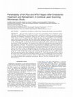
The aim of the study was to assess the penetrability of two endodontic sealers (AH Plus and MTA F... more The aim of the study was to assess the penetrability of two endodontic sealers (AH Plus and MTA Fillapex) into dentinal tubules, submitted to endodontic treatment and subsequently to endodontic retreatment. Thirty ex vivo incisors were prepared using ProTaper rotary system up to F3 instrument and divided in three groups according to the endodontic sealer used for root canal filling: AH Plus (AHP), MTA Fillapex (MTAF), and control group (CG) without using EDTA previously to the root canal filling. Rhodamine B dye (red) was incorporated to the sealers in order to provide the fluorescence which will enable confocal laser scanning microscopy (CLSM) assessment. All specimens were filled with gutta-percha cones using the lateral compaction technique. The specimens were submitted to endodontic retreatment using Pro-Taper Retreatment system, re-prepared up to F5 instruments and filled with gutta-percha cones and the same sealer used during endodontic retreatment. Fluorescein dye (green) was incorporated to the sealer in order to distinguish from the first filling. The roots were sectioned 2 mm from the apex and assessed by CLSM. No difference was found between the two experimental groups (P > 0.05). On the other hand, in the control group the sealers were not capable to penetrate into dentinal tubules after endodontic treatment (P > 0.05). In retreatment cases, none of the sealers were able to penetrate into dentin tubules. It can be concluded that sealer penetrability is high during endodontic treatment. However, MTA Fillapex and AH Plus do not penetrate into dentinal tubules after endodontic retreatment. Microsc. Res. Tech. 00:000-000, 2014.
Influence of technique and manipulation on self-adhesive resin cements used to cement intraradicular posts
The Journal of Prosthetic Dentistry, 2013
Resin cements are widely used to cement intraradicular posts, but bond strength is significantly ... more Resin cements are widely used to cement intraradicular posts, but bond strength is significantly influenced by the technique and material used for cementation. The purpose of this study was to evaluate the bond strength of 3 self-adhesive cements used to cement intraradicular glass fiber posts. The cements all required different application and handling techniques. Forty-five human maxillary canines were selected and randomly divided into 3 groups n= 15 by drawing lots: Group BIS - Biscem, Group BRE - Breeze, and Group MAX - Maxcem. Each group was divided into 3 subgroups according to application and handling techniques: Sub-group A - Automix/Point tip applicator, Sub-group L - Handmix/Lentulo, and Sub-group C - Handmix/Centrix. Cementation of the posts was performed according to the manufacturers&amp;amp;amp;amp;amp;amp;amp;amp;amp;amp;amp;amp;amp;amp;amp;amp;amp;amp;amp;amp;amp;amp;amp;amp;amp;amp;amp;amp;amp;amp;amp;amp;amp;amp;amp;amp;amp;amp;amp;amp;amp;amp;amp;amp;amp;amp;amp;amp;amp;amp;amp;amp;amp;amp;amp;amp;amp;amp;amp;amp;amp;amp;amp;amp;amp;amp;amp;amp;amp;amp;amp;amp;amp;amp;#39; instructions. The push-out test was performed with a crosshead speed of 0.5 mm/min, and bond strength was expressed in megapascals. The results were evaluated by 2-way ANOVA and the all pairwise multiple comparison procedures (Tukey test) (α=.05). Breeze cement showed the highest average for the subgroups A, L, and C when compared to the Biscem cement and Maxcem Elite (P&amp;amp;amp;amp;amp;amp;amp;amp;amp;amp;amp;amp;amp;amp;amp;amp;amp;amp;amp;amp;amp;amp;amp;amp;amp;amp;amp;amp;amp;amp;amp;amp;amp;amp;amp;amp;amp;amp;amp;amp;amp;amp;amp;amp;amp;amp;amp;amp;amp;amp;amp;amp;amp;amp;amp;amp;amp;amp;amp;amp;amp;amp;amp;amp;amp;amp;amp;amp;amp;amp;amp;amp;amp;amp;lt;.05). Statistically significant differences among the subgroups were only observed for Biscem. This study shows that application and handling techniques may influence the bond strength of different self-adhesive cements when used for intraradicular post cementation.
Effect of mechanical loading on the removal torque of different types of tapered connection abutments for dental implants
The Journal of Prosthetic Dentistry, 2013
The mechanical behavior of internal taper implant abutment designs needs to be evaluated. The pur... more The mechanical behavior of internal taper implant abutment designs needs to be evaluated. The purpose of this study was to evaluate the effect of simulated mechanical loading on the removal torque of 1-piece and 2-piece abutments connected to internal taper oral implants. Forty-eight internally notched taper implants were divided into 2 groups of 24. Group OP received solid (1-piece) abutments; group TP received esthetic (2-piece) abutments. Each group was further subdivided into subgroups C (control) without mechanical loading and T (test) with mechanical loading. In groups OPC and TPC, the abutments were placed and removed and the removal torque values (RTVs) registered. In groups OPT and TPT, abutments were placed, mechanically loaded (500 000 cycles), removed, and the RTVs registered. Groups TPC and TPT were further tested for the traction force necessary to dislodge the abutment from the implant. For data analysis, the Student t test (for RTVs) and the Mann-Whitney U test (for TFVs) (α=.05) were performed. All abutments tested presented torque loss with RTVs lower than the placement torque. A statistically significant difference (P=.002) was found between groups OPC (81.6% of placement torque) and OPT mean RTVs results (85.0% of placement torque), while no statistical differences (P=.362) were found between groups TPC (63.7% of placement torque) and TPT (59.1% of placement torque). The traction force values necessary to dislodge the abutment from the implant, however, were significantly higher (P&amp;amp;amp;amp;amp;amp;lt;.001) for group TPT than for group TPC. Cold welding did not occur in any of the abutment specimens tested. Even after the mechanical loading, esthetic abutments presented similar RTVs. The traction force necessary to remove esthetic abutments from inside the implants presented a 2-fold increase after mechanical loading.
Effect of cavity design on tooth surface strain
The Journal of Prosthetic Dentistry, 2013
The loss of tooth structure can increase cuspal flexure, thereby reducing the fracture resistance... more The loss of tooth structure can increase cuspal flexure, thereby reducing the fracture resistance of the tooth, or open the tooth-restoration interface, leading to microleakage. The purpose of this study was to evaluate tooth strain in teeth with different cavity preparations after loading and unloading. Ten intact human maxillary premolars were selected and embedded in epoxy resin molds. Constantan strain gauges were used and tested as an intact tooth (group I), occlusal cavity (group O), mesio-occlusal cavity (group MO), and finally mesio-occluso-distal cavity (group MOD). All teeth were subjected to gradual nondestructive occlusal loading and unloading (50 N, 70 N, 90 N, 110 N, 130 N, 50 N, 0 N) in a servohydraulic testing machine. All data were analyzed statistically by performing a repeated measures ANOVA with load and cavity as factors to compare the relevant mean strains, and a Bonferroni post hoc test was performed for multiple comparisons (α=.05). The repeated measures ANOVA did not provide any evidence of an interaction between load and cavity but indicated a significant difference in the mean strains both between the loads (P&amp;amp;amp;amp;amp;amp;amp;amp;amp;amp;amp;amp;amp;amp;amp;amp;amp;amp;amp;amp;amp;amp;amp;amp;lt;.001) and between the cavity groups (P&amp;amp;amp;amp;amp;amp;amp;amp;amp;amp;amp;amp;amp;amp;amp;amp;amp;amp;amp;amp;amp;amp;amp;amp;lt;.001). MOD cavities presented statistically significantly higher values of strain than MO, O, or intact teeth, and a significant increase in the values of mean strain for all cavities was observed, even with intact teeth, when nondestructive occlusal loading was increased.
Effect of a crown ferrule on the fracture resistance of endodontically treated teeth restored with prefabricated posts
The Journal of Prosthetic Dentistry, 2006
Root fracture is one of the most serious complications following restoration of endodontically tr... more Root fracture is one of the most serious complications following restoration of endodontically treated teeth. The purpose of this study was to compare the fracture strengths of endodontically treated teeth using posts and cores and variable quantities of coronal dentin located apical to core foundations with corresponding ferrule designs incorporated into cast restorations. Fifty freshly extracted canines were endodontically treated. The teeth were randomly divided into groups of 10 and prepared according to 5 experimental protocols. teeth with custom cast post and core; 0-mm group: teeth without coronal structure (no ferrule); 1-mm, 2-mm, and 3-mm groups: teeth with 1 mm, 2 mm, and 3 mm of remaining coronal tooth structure (1-, 2-, and 3-mm ferrule), respectively. All specimens in 0-mm through 3-mm (noncontrol) groups were restored with a prefabricated post (Screw-Post) and composite resin (Z100) core located superior to the different tooth structure heights. All teeth were restored with complete metal crowns. The fracture resistance (N) was measured in a universal testing machine at 45 degrees to the long axis of the tooth until failure. Data were analyzed by 1-way analysis of variance and Tukey test (alpha=.05). Significant differences (P&amp;amp;amp;amp;amp;amp;amp;amp;amp;amp;lt;.001) were found among the mean fracture forces of the test groups (control group: 818.2 N; 0-mm, 1-mm, 2-mm, and 3-mm groups: 561.0 N, 627.6 N, 745.3 N, and 907.1 N, respectively). When the mode of failure was evaluated, all failures in the control group occurred due to root fracture, and all failures in the 0-mm group occurred due to core fracture. The majority of failures in the other groups occurred due to crown cementation failure. The results of this study showed that an increased amount of coronal dentin significantly increases the fracture resistance of endodontically treated teeth.
The Journal of Prosthetic Dentistry, 2013
Pereira JR, Valle AL, Ghizoni JS, S o MVR, Ramos MB, Lorenzoni FC. Evaluation of push-out bond st... more Pereira JR, Valle AL, Ghizoni JS, S o MVR, Ramos MB, Lorenzoni FC. Evaluation of push-out bond strength of four luting agents and SEM observation of the dentine/ fibreglass bond interface. International Endodontic Journal.
Fracture resistance of endodontically treated teeth restored with glass fiber posts of different lengths
The Journal of Prosthetic Dentistry, 2014
Endodontically treated teeth are known to have reduced structural strength. Glass fiber posts may... more Endodontically treated teeth are known to have reduced structural strength. Glass fiber posts may influence fracture resistance and should be evaluated. The purpose of this study was to evaluate the influence of glass fiber post length on the fracture resistance of endodontically treated teeth. Forty intact human maxillary canines were selected and divided into 4 groups, the control group consisting of teeth restored with a custom gold cast post and core, with a length of two-thirds of the root. Other groups received prefabricated glass fiber posts in different lengths: group 1/3, removal of one-third of the sealing material (5 mm); group 1/2, removal of one-half of the sealing material (7.5 mm); and group 2/3, removal of two-thirds of the sealing material (10 mm). All the posts were cemented with resin cement, and the specimens with glass fiber posts received a composite resin core. All the specimens were restored with a metal crown and submitted to a compressive load until failure occurred. The results were evaluated by 1-way ANOVA, and the all pairwise multiple comparison procedures (Tukey honestly significantly difference test) (α=.05). The ANOVA showed significant differences among the groups (P&amp;amp;amp;amp;lt;.002). The Tukey test showed that the control group presented significantly higher resistance to static load than the other groups (control group, 634.94 N; group 1/3, 200.01 N; group 1/2, 212.17 N; and group 2/3, 236.08 N). Although teeth restored with a cast post and core supported a higher compressive load, all of them fractured in a catastrophic manner. For teeth restored with glass fiber posts, the failure occurred at the junction between the composite resin core and the root. The length of glass fiber posts did not influence fracture load, but cast post and cores that extended two-thirds of the root length had significantly greater fracture resistance than glass fiber posts.

Journal of Applied Oral Science, 2005
RESUMO www.fob.usp.br/revista or www.scielo.br/jaos bjectives: The aim of this study was to evalu... more RESUMO www.fob.usp.br/revista or www.scielo.br/jaos bjectives: The aim of this study was to evaluate the influence of remaining coronal tooth structure on endodontically treated teeth restored with prefabricated posts and two different composites for core build-up: dual-cured resin (Enforce Core) and light-cured resin (Z-250). Methods: Fourty freshly extracted canines were endodontically treated and divided into four groups: Group I -teeth with 3mm remaining coronal structure, restored with Enforce Core; Group II -teeth with 3mm remaining coronal structure, restored with Z-250; Group III -teeth with no remaining coronal structure, restored with Enforce; Group IV -teeth with no remaining coronal structure, restored with Z-250. After restoration, the teeth were embedded in acrylic resin and the fracture resistance was measured on a universal testing machine at 45 degrees to the long axis of the tooth until failure. Results: Data were analyzed by two-way analysis of variance, which showed significant differences between groups (p=0.00). The Tukey test did not show significant differences between specimens with and without remaining coronal structure. Conversely, significant difference was observed between groups with different core build-up. The highest values of fracture resistance were found in the group restored with light-cured resin. Significance: The remaining coronal tooth structure did not influence the resistance of endodontically treated teeth; however, the change of core build-up was able to modify this resistence. Uniterms: Post and core technique; Composite resins; Fracture strength. objetivo desta pesquisa foi avaliar a influência do remanescente dentário coronal de dentes tratados endodonticamente, restaurados com pinos pré-fabricados e duas resinas como núcleos de preenchimento, uma de presa dual (Enforce Core) e outra fotopolimerizável (Z-250). Foram utilizados 40 caninos superiores humanos extraídos, divididos em quatro grupos de 10 espécimes: Grupo l -com remanescente dentário coronal de 3mm e restaurados com Enforce Core; Grupo ll -com remanescente dentário coronal de 3mm e restaurado com Z-250; Grupo III -sem remanescente dentário coronal e restaurado com Enforce Core; Grupo IV -sem remanescente dentário coronal e restaurado com Z-250. Após restaurados, os dentes foram levados a uma Máquina de Ensaio Universal e submetidos a uma força de compressão à 45º até que ocorresse fratura da restauração. A análise dos resultados (ANOVA, p>0,05) mostrou não haver diferença estatisticamente significativa entre os dentes com e sem remanescente dentário coronal. Com relação ao material utilizado para o preenchimento coronário, constatou-se diferença significativa, sendo que os valores mais elevados de resistência à fratura foram encontrados no grupo restaurado com a resina fotopolimerizável. Unitermos: Pinos de retenção dentária; Resinas compostas; Resistência a fratura.
Journal of Applied Oral Science, 2007

Uploads
Papers by jefferson pereira